A tissue expression atlas is a reference set that measures, for every protein-coding gene, how much of it is found in each human organ, tissue or cell type — so that a search on one gene symbol returns an organ-by-organ pattern rather than a single number. For a toxicologist the atlas is not the answer; it is the map that decides which questions get asked. Where the target sits tells you which organs a liability list has to cover, and whether the species your four-week study runs in shares the pattern at all. Botminds' Target Safety Assessment runs expression as one of six evidence lines, summarising where the target is in human tissues and cells and in mouse, rat, dog and monkey, and flagging the species where the pattern differs.
Examples from the product on this page use sample data on fictional targets, people and companies; the research figures come from regulators and peer-reviewed studies, linked where they appear.
What the public atlases now cover
44tissues
human tissues in the reference set behind the genetic effects on expression described by the GTEx Consortium
192clusters
individual cell type clusters in the open-access single-cell map of human tissues
15tissues
deep-sequenced in both human and mouse in a comparison whose species-level headline was later disputed
What is a tissue expression atlas, and what is in one?
An atlas is a measurement, repeated across tissues with one method, so that the numbers can be compared with each other. Three kinds sit behind most target work. Bulk RNA atlases sequence whole tissue samples: the GTEx Consortium's analysis of genetic effects on gene expression levels across 44 human tissues is the largest of them, and its value to a toxicologist is less the genetics than the breadth — donors, organs and replicates in one normalised set. Antibody-based atlases stain tissue sections and report protein, at the cost of depending on one antibody's specificity. Single-cell atlases break the tissue apart first and report expression per cell type; the open-access map published in Science Advances lets a reader explore the expression of human protein-coding genes in 192 individual cell type clusters.
What an atlas does not contain matters as much. It is a healthy-tissue reference, usually from adults, usually at rest. It says nothing about what happens to expression under inflammation, injury or drug pressure, nothing about the fetus unless a developmental atlas was used, and — unless the atlas is explicitly cross-species — nothing about the rat. Every one of those gaps is a place where an assessment can be wrong while every cited number is right.
From an expression pattern to a list of organs at risk
The reasoning from expression to liability is short and worth writing out, because it is where most of the value is. A drug that engages its target does so wherever the target is. So the organs where the target is abundant are the organs where on-target pharmacology will happen, and the question for each of them is whether that pharmacology is tolerable. Expression does not score a risk by itself — it decides which organs have to be asked about and which can be left clear.
The published target safety assessment of plasmepsin IX/X, written for an antimalarial programme, shows the move in two sentences. Having found that the nearest human relatives of the parasite enzyme are the aspartic proteases cathepsin D and E, the authors reason from where those enzymes sit to what could be hurt: Of these, CatD/E may be of most toxicological relevance as CatD is a ubiquitous lysosomal enzyme present in most cell types and CatE is found in the gut and in erythrocytes, the clinically significant site of malarial infection
(Barber et al., Toxicology Research, 2021). One enzyme is everywhere, so the liability is general and lysosomal; the other is in two places, so the liability is named: the gut, and red cells. That is an expression line doing its job.
The expression line of an assessment: the fields, and what a missing one costs
| Field | What it has to say | What goes wrong when it is left out |
|---|---|---|
| Organ system | The organ or tissue the measurement belongs to, in the same list the liability register uses. | A finding that cannot be filed against an organ cannot be weighed against the knockout or the carrier data for that organ. |
| Species | Human, and each toxicology species the pattern was checked in. | A human-only line is read as though it settled the species question, and the study is designed in an animal that may not express the target. |
| RNA or protein | Which was measured. Never both in one cell. | A transcript count is quoted as though a protein had been seen, and a tissue with message but no detectable protein is scored as a target organ. |
| Level and cell type | How much, and in which cells — epithelium, myeloid cells, endothelium — not only which organ. | A whole organ is flagged on a signal that belongs to one rare cell type in it, or a rare cell type is lost inside a whole-organ average. |
| Source passage | The atlas entry or paper passage the value came from, quotable as written. | The number cannot be re-checked when the atlas is updated, and a reviewer has to take it on trust. |
| Species difference | An explicit note where the animal pattern is not the human one. | The disagreement surfaces in the study report instead of in the assessment, after the money is spent. |
Does low expression in a healthy tissue matter?
Yes, and the clearest evidence is a clinical case. A patient treated with T cells engineered against ERBB2 for metastatic colon cancer deteriorated within minutes of the infusion and died five days later. The investigators' own reading of the event was that the transferred cells lodged in the lung and were triggered to release cytokine by recognising the low levels of the same protein on normal lung epithelial cells (Morgan et al., Molecular Therapy, 2010). The lung was not a target organ by any ordinary reading of an expression table. It was a target organ for a drug potent enough to respond to a small signal.
The lesson is not that every low value is dangerous; most are not. It is that "low" is a statement about the measurement, and risk is a statement about the measurement and the molecule together. A highly potent, long-acting or cell-based agent converts a weak signal into a real exposure; a modest small molecule with a short half-life may not. So an assessment should never write "not expressed" when it means "below the atlas's detection threshold", and should carry the modality beside the pattern.
Two conclusions a toxicologist can draw from the same low-expression finding
The reading that closes the question: Target not expressed in lung — no pulmonary liability.
The reading that keeps it open: Target detected in lung epithelium at low level, RNA only, one atlas, no protein measurement.
The reading that closes the question: That a value below a reporting threshold is a zero, and that a zero in healthy tissue is a zero under treatment.
The reading that keeps it open: That a threshold is a property of the assay, and that potency decides what a small amount of target is worth.
The reading that closes the question: A potent or long-resident agent finds the protein the table rounded away.
The reading that keeps it open: It does not close; the organ stays on the list until a protein measurement or a study removes it.
The reading that closes the question: Nothing, until it costs everything — the published cell-therapy case turned on recognition of low levels on lung epithelium.
The reading that keeps it open: One line in the monitoring plan, or one stain.
RNA and protein are two findings, not one
Transcript abundance and protein abundance are measured by different assays, correlate imperfectly, and answer different questions. A drug binds protein. An atlas that reports transcripts is evidence that protein could be there, not that it is — and the gap between the two is largest exactly where it matters: tissues with high turnover, secreted proteins, and genes under strong post-transcriptional control.
This is why the expression agent in Target Safety Assessment carries one explicit limit: it reports RNA and protein separately, and never merges them into a single "expressed" verdict. The discipline costs a column and buys the ability to say what kind of evidence an organ is on. A liability supported by protein staining in the target cell type is a different claim from one supported by a transcript count in whole tissue, and a reviewer who cannot tell them apart cannot weigh the organ.
Two habits follow. Write the assay, not the conclusion: "RNA, whole colon, high" rather than "expressed in colon". And when the two disagree — message present, protein absent, or the reverse — record the disagreement rather than resolving it silently, in the same way a mouse finding and a human finding that disagree are both kept. Which evidence line wins is a judgement, and judgements belong to the toxicologist who signs.
How similar is mouse and human tissue expression?
The honest answer is that it depends on the gene, and the most quoted genome-wide answer is contested. A deep RNA-sequencing comparison of 15 tissues in both species, published in PNAS, reported that in this study of a broad number of tissues between humans and mice, high-throughput sequencing assays on the transcriptome and epigenome reveal that, in general, differences dominate similarities between the two species
. The sharper finding is in the next sentence: although commonalities are evident in the expression of tissue-specific genes between the two species, the expression for many sets of genes was found to be more similar in different tissues within the same species than between species
(Lin et al., PNAS, 2014). A reanalysis of the same data then argued that the study design had confounded species with sequencing batch, and that once the batch effect was accounted for, the human and mouse samples clustered by tissue rather than by species (Gilad and Mizrahi-Man, F1000Research, 2015).
For a target assessment the dispute is itself the lesson. A genome-wide average, in either direction, says little about the one gene in front of you: tissue-specific genes held their patterns even in the original analysis, and a single gene may be perfectly conserved in its distribution or may not. What neither paper licenses is the default assumption: that because a gene is present in the mouse, the organ it sits in is the same organ it sits in here. The assumption has to be checked for the one target in front of you, and the check has to be recorded, because the species the toxicology study runs in is chosen from it.
Expression as one line of evidence, filed against the organ it belongs to
How do you choose the toxicology species for a target?
For a biological medicine the regulator answers this directly, and the answer is expression. ICH S6(R1) defines the test in one sentence, and it is worth quoting because it is narrower than the way the question is usually discussed — it is about pharmacology, not about taxonomy or convenience.
A relevant species is one in which the test material is pharmacologically active due to the expression of the receptor or an epitope (in the case of monoclonal antibodies).
ICH S6(R1), Preclinical Safety Evaluation of Biotechnology-Derived Pharmaceuticals, 2011
For antibodies the guideline adds a second requirement that is purely an expression question: relevant species are those that express the desired epitope and show a tissue cross-reactivity profile similar to human tissues. Tissue cross-reactivity is the study that asks it — the labelled antibody is applied to panels of human and animal tissues to see what it binds, wanted or not. An assessment that has already written down where the target is, in which species, and whether the pattern matches, has done most of the reading that study design depends on.
Small molecules are not covered by that definition, and the species choice there turns on metabolism and on the pharmacology of the target in each species. But the discipline transfers: if the organ you are worried about does not carry the target in the species you dose, the study cannot see the effect you are afraid of, and a clean result from it means nothing.
What a species-relevance claim has to carry before a study is designed on it
- The target is expressed in the species, in the organ at issue
Not merely present in the genome: measured in the tissue the liability names, with the assay stated.
- The measurement is comparable to the human one
Same kind of assay where possible. A protein stain in one species against a transcript count in the other is not a comparison.
- For a biological medicine, pharmacological activity is shown, not assumed
Binding, potency or a functional readout in that species — the guideline asks for activity due to expression of the receptor or epitope.
- Any difference in pattern is written as a difference, with the organ named
Flagging "species differ" without saying where leaves the study director nothing to design around.
- The source passage is attached to each value
An atlas entry or a paper passage a reviewer can open, so the claim survives a challenge and an atlas update.
- The modality is stated beside the pattern
A cell therapy, a long-acting antibody and a short-half-life small molecule do not take the same risk from the same low value.
When no species expresses the target
Sometimes the honest answer is that no ordinary toxicology species is relevant: the epitope is human-specific, the receptor has diverged, or the pharmacology simply does not occur in the rat or the dog. The guideline anticipates this and names two routes rather than leaving the programme to improvise — when no relevant species exists, the use of relevant transgenic animals expressing the human receptor or the use of homologous proteins should be considered
.
Both routes change what the study can prove, and an assessment should say so. A transgenic animal expressing the human receptor tests the pharmacology, but in a tissue distribution the transgene decides rather than human biology; a homologous protein — the analogous molecule against the animal's own target — tests the consequence of hitting the pathway, but not the clinical molecule. Neither is a reason to skip the question. Both are reasons to write the limitation into the assessment at target stage, where it is a paragraph, rather than at the pre-trial meeting, where it is a delay.
Single-cell resolution, and what it changes
Whole-tissue measurement averages across everything in the sample. A gene expressed intensely in a cell type that makes up one per cent of an organ looks low; a gene spread thinly through the bulk looks the same. Single-cell atlases separate those two cases, and for safety work the distinction is often the whole finding: what is at risk is the cell type, not the organ.
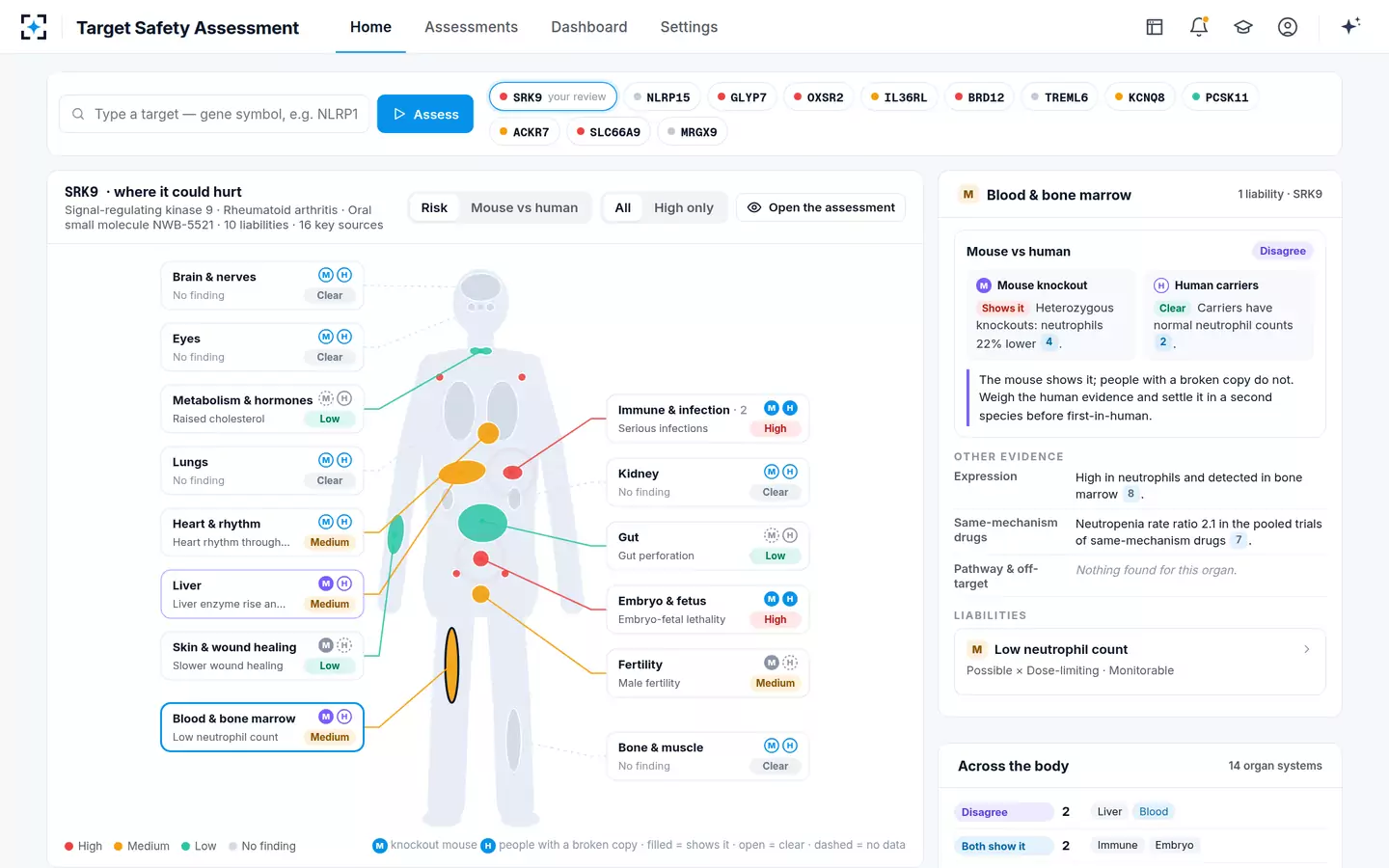
In the sample assessment on this page, the fictional target is described as high in colonic epithelium and the knockout animals get worse chemically induced colitis — a cell type and a consequence that line up. The same target shows low expression in heart muscle, which is why the cardiac question on it is settled by other evidence rather than by the expression line; that argument is worked through in the guide to settling a mouse QT finding. Cell-type resolution does not make expression decisive. It makes the claim specific enough to be argued with.
Which expression source should an assessment use?
Whichever ones you can cite, in that order of preference: protein in the human tissue at issue, then human transcript data from a normalised multi-tissue set, then animal data of the same kind, then anything else, labelled. Mixing atlases is normal; pretending they are interchangeable is not, because thresholds, units and normalisation differ between them and a value moved from one to another stops meaning what it meant.
Two practical rules keep this manageable. Name the source per value, not per section, so a reviewer can see that the lung number and the colon number came from different places. And re-read the source when the assessment is re-opened: atlases are versioned, and a tissue that was absent from an older release may be present in the current one. A target assessment that is re-checked after signing — the default in Target Safety Assessment is weekly — inherits that discipline for free.
How Target Safety Assessment records expression by organ and species
The expression agent reads the atlases the team allows and summarises where the target is, by organ system, in human tissues and cells and in mouse, rat, dog and monkey, flagging the species where the pattern differs. Its findings land against the same fourteen organ systems the rest of the assessment uses, so the expression line for the gut sits beside the knockout finding for the gut and the carrier scan for the gut, rather than in a section of its own. Its one stated limit is the one this page has been arguing for: RNA and protein are reported separately.
Expression is one supporting line of six — and rarely the one that decides
Two places in the product turn the expression line into a decision rather than a note. The first is the organ panel, where expression sits under the other evidence for that organ with its cited passage, so a reviewer can see at a glance whether the organ is on the list because of where the target is or because of what the knockout did. The second is the report: species relevance and study design is a section of the house template, switched on in the sample settings, and it is written from the expression line. What the register does with the result is set out in the guide to the studies that settle a liability.
One liability in Target Safety Assessment, with the expression row stating plainly that it has nothing to add
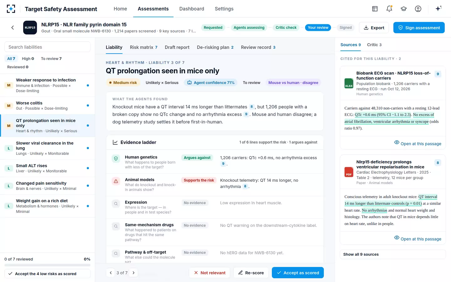
The practical test of all this is whether an assessment can answer, for any organ on its list, three questions in one breath: is the target there, in which species, and measured how. Where the answers differ between the human and the animal, that difference is the sentence that decides the study. To see the expression line against the knockout and the carrier evidence on one body map, start from Target Safety Assessment.
Questions people ask
What is a tissue expression atlas used for in drug safety?
It is used to decide which organs an on-target liability list has to cover, and to check whether the toxicology species shares the human pattern. It does not score risk on its own: a target can be abundant in an organ with no adverse consequence, and scarce in one where the pharmacology is intolerable. The atlas sets the agenda; the knockout, carrier and drug-label evidence settle it.
Is RNA expression the same as protein expression?
No. They are different measurements with imperfect correlation, and a drug binds protein. Transcript data in a tissue is evidence that protein may be present there, and a reasonable basis for asking the question; it is not evidence that the protein is present at a level a drug will engage. An assessment should say which was measured for every value it stores.
How do you choose the toxicology species for a biological medicine?
By pharmacological activity, which in practice means expression. ICH S6(R1) defines a relevant species as one in which the test material is pharmacologically active because the receptor or epitope is expressed, and for monoclonal antibodies adds that the species should show a tissue cross-reactivity profile similar to human tissues. Where no relevant species exists, the guideline points to transgenic animals expressing the human receptor, or to a homologous protein.
What is tissue cross-reactivity?
It is a study in which a labelled antibody is applied to panels of human and animal tissue sections to see everything it binds — the intended target and anything else. It supports species selection by showing whether the animal's binding pattern resembles the human one, and it can reveal binding in tissues no expression table predicted. It is an immunohistochemical screen, so it shows where binding occurs rather than what the binding does.
Does a target have to be expressed in a tissue for that tissue to be damaged?
No. On-target damage follows expression, but an organ can be harmed downstream of pharmacology elsewhere — through a mediator, a haemodynamic change or a metabolic consequence — and off-target binding does not follow the target's pattern at all. Expression tells you where on-target effects can occur; it does not bound the list of organs that can be affected.
How different are mouse and human tissue expression?
It varies by gene, and the genome-wide answer is contested. A 15-tissue RNA sequencing comparison reported that differences generally dominate similarities between the species; a later reanalysis attributed that pattern to a confound between species and sequencing batch, and found the corrected data clustering by tissue. Either way, the mouse pattern for one target has to be looked up rather than assumed.